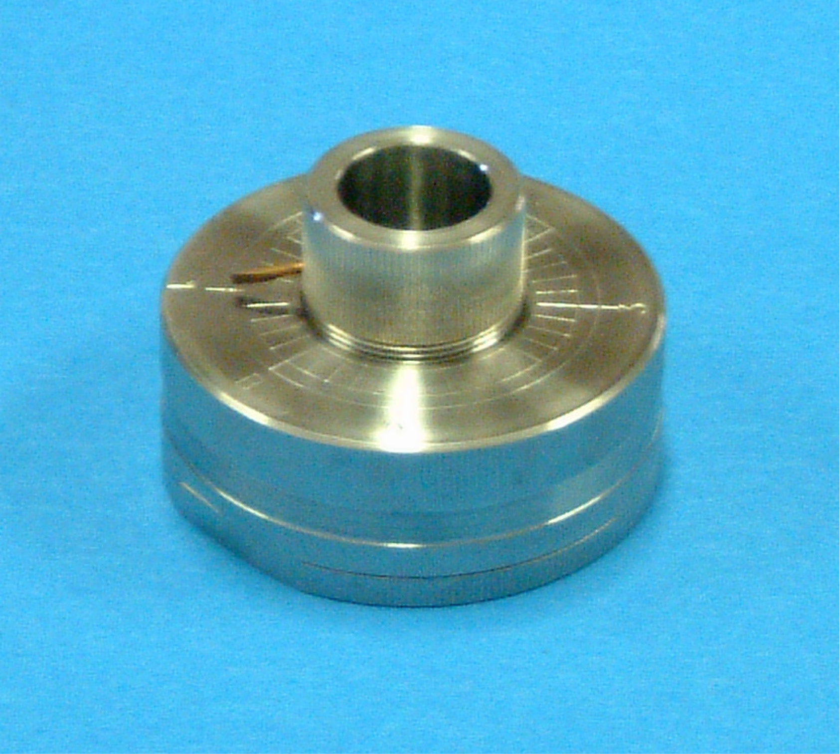
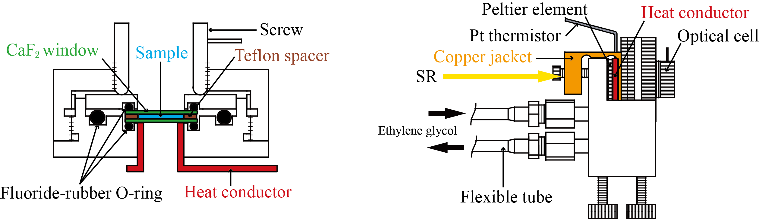
HiSOR SR-VUVCD


Japanese

[SR-VUVCD spectrophotometer]
Optical device
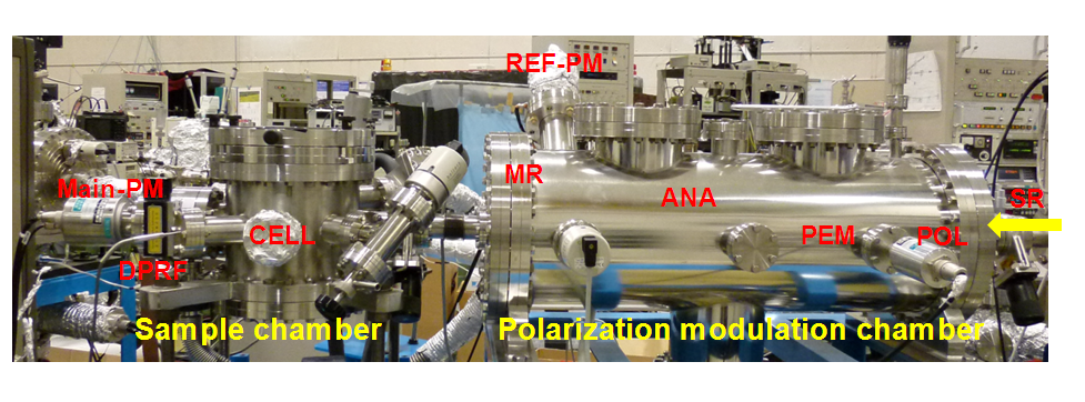
 SR-VUVCD spectrophotometer consists of two separate vacuum chambers (at10-4 Pa; a polarization modulation chamber and a sample chamber). All optical devices of the spectrophotometer are set up under a high vacuum (10-6 Torr), in order to avoid the light absorption in the VUV region by air and water vapor. The SR light is separated into two orthogonal linearly polarized light beams by a linear polarizer (POL, MgF2). Both linearly polarized light beams are modulated into circularly polarized light at 50 kHz by a photo-elastic modulator (PEM, LiF). The main light beam is led to the sample cell and the CD signal is detected by a photomultiplier tube (Main-PM), which is covered by an MgF2 window. The CD signal is rectified and amplified by the lock-in amplifier (LIA), and finally recorded on a personal computer. Another light beam from the PEM is detected as a reference signal by another photomultiplier tube (Ref-PM) after being modulated into a linearly polarization light beam at 100 kHz by a linear polarizer (ANA, MgF2), to obtain optimal driving voltages for the PEM. Further, this reference signal is used to enhance the stabilization of the LIA through the synchronization and rectification of the CD signal detected by the Main-PM.
Picture and Block diagram of SR-VUVCD
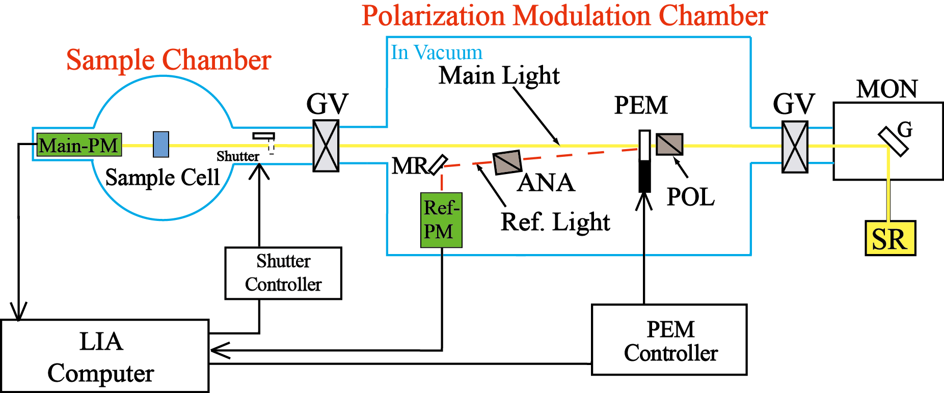
ANA, analyzer; G, grating; GV, gate valve; LIA, lock-in amplifier; MON, monochromator; MR, mirror; PEM, photoelastic modulator; PM, photomultiplier tube; POL, polarizer; S, shutter; SR, synchrotron radiation.

Sample cell
 Sample cell consists of a stainless-steel container with a cylindrical screw, two CaF2 (or MgF2) crystal windows of 20-mm diameter and 1-mm thickness, and three fluoride-rubber O-rings. The optical path length can be adjusted over the range from 1.3 μm (without a spacer) to 100 μm (with donut-shaped Teflon spacers). The sample solution in the cell is sealed with three fluoride-rubber O-rings. The temperature of the sample is controlled between –20°C and 70°C by a Peltier thermoelectric element fixed to the copper jacket in which coolant from a thermostatic bath is circulated through flexible stainless-steel tubes.
       Picture of optical cell                       BLock diagrams of optical cell and temperature control unit


[Applications to structural analysis of biomolecules]

Structural analysis of sugars
 D-Glucose and D-mannose, and D-galactose exhibit markedly different VUVCD spectra despite having very similar structures. The spectra of D-glucose and D-mannose show positive peaks around 168 and 170 nm, respectively. In contrast to these two monosaccharides, D-galactose shows two negative CD peaks around 177 and 162 nm. The spectra in the range 160–180 nm can be assigned to the n-s* transition of lone-pair oxygen electrons of hydroxy groups and acetal bonds, based on simple quantum mechanical considerations at the molecular orbital level. These VUVCD spectra were significantly affected by the a- and β-anomers at C-1, and the trans (T) and gauche (G) configurations of hydroxymethyl group at C-5.
  UVCD spectra of D-glucose, D-mannose, and D-galactose                        Component spectra of D-glucose

Structural analysis of proteins
VUVCD spectra of proteins sensitively reflect secondary structures such as a-helices, β-sheets, turns, and unordered structures. Therefore, we can estimate the contents and the numbers of segments of these secondary structures of an unknown protein from its VUVCD spectra using some analytical programs (e.g. SELCOM 3 program). These secondary-structure data can be further combined with bioinformatics to improve the sequence-based predictions (VUVCD-NN method). The scheme for the secondary-structure analysis of proteins by VUVCD spectroscopy is depicted in the following illustration.
Scheme for the secondary-structure analysis of proteins by SR-VUVCD spectroscopy

[References]

1)      N. Ojima, K. Sakai, K. Matsuo, T. Matsui, T. Fukazawa, H. Namatame, M. Taniguchi, and K. Gekko (2001) “Vacuum-ultraviolet circular dichroism spectrophotometer using synchrotron radiation: optical system and on-line performance”, Chem. Lett., 522-523.

2)      K. Matsuo, Y. Matsushima, T. Fukuyama, S. Senba, and K. Gekko (2002) “Vacuum-ultraviolet circular dichroism of amino acids as revealed by synchrotron radiation spectrophotometer”, Chem. Lett., 826-827.

3)      K. Matsuo, K. Sakai, Y. Matsushima, T. Fukuyama, and K. Gekko (2003) “Optical cell with a temperature-control unit for a vacuum-ultraviolet circular dichroism spectrophotometer”, Analytical Science, 19, 129-132.

4)      K. Matsuo and K. Gekko (2004) “Vacuum-ultraviolet circular dichroism study of saccharides by synchrotron radiation spectrophotometry”, Carbohydrate Research, 339, 591-597.

5)      K. Matsuo, R. Yonehara, and K. Gekko (2004) “Secondary-structure analysis of proteins by vacuum-ultraviolet circular dichroism spectroscopy”, J. Biochem., 135, 405-411.

6)      K. Matsuo, T. Fukuyama, R. Yonehara, H. Namatame, M. Taniguchi, and K. Gekko (2005) “Vacuum-ultraviolet circular dichroism spectrophotometer using synchrotron radiation”, Journal of Electron Spectroscopy and Related Phenomena, 144-147, 295–297.

7)      K. Gekko, R. Yonehara, Y. Sakurada, and K. Matsuo (2005) “Structure analyses of biomolecules using a synchrotron radiation circular dichroism spectrophotometer”, Journal of Electron Spectroscopy and Related Phenomena, 144–147, 1023–1025.

8)      K. Matsuo, R. Yonehara, and K. Gekko (2005) “Improved estimation of the secondary structure of protein by vacuum-ultraviolet circular dichroism spectroscopy”, J. Biochem. 138, 79–88.

9)      T. Fukuyama, K. Matsuo, and K. Gekko (2005) “Vacuum-ultraviolet electronic circular dichroism of L-alanine in aqueous solution investigated by time-dependent density functional theory”, J. Phys. Chem.A 109, 6928–6933.

10)  K. Gekko and K. Matsuo (2006) “Vacuum-ultraviolet circular dichroism analysis of biomolecules”, Chirality, 18, 329–334.

11)  K. Matsuo, Y. Sakurada, R. Yonehara, M. Kataoka, and K. Gekko (2007) “Secondary-Structure Analysis of Denatured Proteins by Vacuum-Ultraviolet Circular Dichroism Spectroscopy”Biophys. J., 92, 4088–4096.

12)  K. Matsuo, H. Watanabe, K. Gekko (2008)“Improved sequence-based prediction of protein secondary structures by combining vacuum-ultraviolet circular dichroism spectroscopy with neural network”Proteins 73, 104–112.

13)K. Matsuo, H. Namatame, M. Taniguchi, K. Gekko (2009) “Vacuum-ultraviolet circular dichroism analysis of glycosaminoglycans by synchrotron-radiation spectroscopy”, Biosci. Biotech. Biochem. 73, 557–561.

14)K. Matsuo, H. Watanabe, S. Tate, H. Tachibana, K. Gekko (2009) ”Comprehensive secondary-structure analysis of disulfide variants of lysozyme by synchrotron-radiation vacuum-ultraviolet circular dichroism”, Proteins, 77, 191–201.

15)  K. Matsuo, H. Namatame, M. Taniguchi, K. Gekko (2009) ”Membrane-induced conformational change of α1-acid glycoprotein characterized by vacuum-ultraviolet circular dichroism spectroscopy”, Biochemistry, 48, 9103–9111.

16)  H. Hiramatsu, M. Lu, K. Matsuo, K. Gekko, Y. Goto, T. Kitagawa (2010) ”Differences in the molecular structure of β2-microglobulin between two morphologically different amyloid fibrils”, Biochemistry, 49, 742–751.

17) T. Fukuyama, K. Matsuo, K. Gekko (2011) ”Experimental and Theoretical Studies of Vacuum-Ultraviolet Electronic Circular Dichroism of Hydroxy Acids in Aqueous Solution”, Chirality, 23, E52-E58.

18)  M. Yagi-Utsumi, K. Matsuo, K. Yanagisawa, K. Gekko, K. Kato (2011) “Spectroscopic Characterization of Intermolecular Interaction of Amyloid β Promoted on GM1 Micelles”, Int. J. Alzheimers Dis., doi:10.4061/2011/925073.

19)  K. Matsuo, Y. Sakurada, S. Tate, H. Namatame, M. Taniguchi, K. Gekko (2012) “Secondary-structure analysis of alcohol-denatured proteins by vacuum-ultraviolet circular dichroism spectroscopy”, Proteins, 80, 281–293.

20)  K. Matsuo, H. Namatame, M. Taniguchi, K. Gekko (2012) “Vacuum-Ultraviolet Electronic Circular Dichroism Study of Methyl α-d-Glucopyranoside in Aqueous Solution by Time-Dependent Density Functional Theory”, J. Phys. Chem. A, 40, 9996–10003.

21)  K. Matsuo, K. Gekko (2013) “Circular-Dichroism and Synchrotron-Radiation Circular-Dichroism Spectroscopy as Tools to Monitor Protein Structurein a Lipid Environment”, Methods Mol. Biol., 974, 151–176.

22)  E. Ohmae, K. Matsuo, K. Gekko (2013) “Vacuum-ultraviolet circular dichroism of Escherichia coli dihydrofolate reductase: Insight into the contribution of tryptophan residues”, Chem. Phys. Lett., 572, 111–114.

23) H. Takekawa, K. Tanaka, E. Fukushi, K. Matsuo, T. Nehira, M. Hashimoto (2013) “Roussoellols A and B, Tetracyclic Fusicoccanes from Roussoella hysterioides, J. Nat. Prod., 76, 1047–1051.

24) K. Matsuo, H. Hiramatsu, K. Gekko, H. Namatame, M. Taniguchi, R. W. Woody (2013) “Construction of a Synchrotron-Radiation Vacuum-Ultraviolet Circular-Dichroism Spectrophotometer and Its Application to the Structural Analysis of Biomolecules, Bull. Chem. Soc. Jpn., 86, 675–689.

25) K. Matsuo, H. Hiramatsu, K. Gekko, H. Namatame, M. Taniguchi, R. W. Woody (2014) “Characterization of Intermolecular Structure of β2-Microglobulin Core Fragments in Amyloid Fibrils by Vacuum-Ultraviolet Circular Dichroism Spectroscopy and Circular Dichroism Theory, J. Phys. Chem. B, 118, 2785–2795.

26) K. Matsuo, H. Namatame, M. Taniguchi, K. Gekko (2015) "Solution structures of methyl aldopyranosides revealed by vacuum-ultraviolet electronic circular-dichroism spectroscopy" Biomedical Spectroscopy and Imaging, 4, 269–282.

Download of program PROTPOL

[Shared-use and joint research]

Our SR-VUVCD spectrophotometer is used as the sharing instrument for the domestic and international researchers.

Contact person:

Dr. Koichi Matsuo
Hiroshima Synchrotron Radiation Center, Hiroshima University
2-313 Kagamiyama
Higashi-Hiroshima 739-0046 Japan
E-mail: pika(at)hiroshima-u.ac.jp
Please change from (at) to @.

Tel: +81-82-424-6293

Fax: +81-82-424-6294