Pediatric liver tumor is a disease presents when malignant (cancer) cells
occur in the livers of children. Two typical types of pediatric liver tumor
are as follows:
- Hepatoblastoma: Liver tumors specific to children
which usually occur in children under age 5.
- Hepatoma (Hepatocellular carcinoma): Liver tumors
which can occur in both adults and children of all ages.
These primary hepatic tumors (tumors which occur initially in the liver)
are called pediatric liver tumors. This study group shows guideline treatments
for these tumors. On principle, this study group does not cover the treatment
of metastatic liver cancer (cancers which spread to liver from other original
sites, such as liver metastasis of neuroblastoma) and rhabdomyosarcoma
in liver.
Children who have certain diseases and/or lesions have high incidence of
pediatric liver tumor.
The followings are the risk factors for hepatoblastoma:
- Familial adenomatous polyposis(FAP)
- Beckwith-Wiedemann syndrome
- Extremely low birth weight
The risk factors for hepatoma are as follows.
- Hepatitis B. or C. If mothers infect their children with hepatitis B or
hepatitis C, risks increase.
- Liver damage caused by certain diseases such as biliary cirrhosis or tyrosinemia.
Possible symptoms of pediatric liver tumor are abdominal stiffness, stomachache
and fever.
The larger tumors become, the more symptoms appear. Please contact a doctor
when the child has symptoms such as the following:
- Indolent palpable abdominal tumor, especially in the right upper quadrant
- Abdominal distensions and pain
- Unexplained weight loss
- Persistent fever
- Anorexia
- Precocious puberty (boys)
- Vomiting and nausea
To find and diagnose pediatric liver tumors, liver function and blood tests are performed.
The following test methods and techniques are presently in use.
- Medical history and clinical examination: Holistic examination including
examination of pathological features such as palpable tumors and finding
out the health lifestyle, medical history and records of the patient.
- Tumor marker blood serum test: Blood is tested for the amount of characteristic
substances of pediatric liver tumor (tumor markers) released into the blood
from organs, tissues and tumor cells. Concentration of a-fetoprotein (AFP)
in the blood increases in most cases of pediatric liver tumor. Moreover,
sometimes the concentration of the hormone b-human chorionic gonadotropin
(b-hCG) and the level of cholesterol produced in the liver also increase.
However, immediately after birth, normal babies also show very high levels
of AFP and levels sometimes increase with other cancers or with non-cancerous
diseases (cirrhosis, hepatitis and etc.) To distinguish these, the type
of AFP is sometimes examined.
- Blood examination:
- The number of red blood cells, leukocytes and platelets.
In cases of hepatoblastoma, tumors which make platelets and produce platelet
producing factors show thrombocytosis.
- The amount of hemoglobin (protein which deliver oxygen) in red blood cells.
- The percentage of red blood cells in the blood
sample.
- Liver function test: Estimates liver function with concentration of specific
substances released into blood from the liver. In case of liver tumors,
these values sometimes appear higher than normal because normal hepatocyte
and biliary tract are pressured or injured.
- Abdominal X-ray: The X-ray inspection of abdominal internal organs. X-ray
is a kind of the radiation. By letting it pass through the human body and
irradiate on a film, the size of liver and image of calcification are detected
on the film.
- Ultrasound examination: By reflecting ultrasonic waves on internal tissues
and organs, tumors in the liver are depicted by imagining of their echo.
It also allows examination of the relationship between tumors and blood
vessels, such as hepatic vein or the portal vein, and the relation with
the inferior vena cava or the biliary.
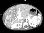
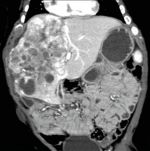
- CT scan (Computed tomography): Photographs the area
inside the body from various angles and creates detailed image sequence. The
images are created by a computer which is connected to an X-ray system. In some
cases, to project organs, tissues, blood vessels or biliary tracts more
clearly, intravenous injections or an oral contrast medium may be needed. In
general, for pediatric liver tumors, CT scans carried out on the chest and abdomen.
Nowadays, we are able to examine the exact location of tumors and relationship
with blood vessels and biliary tracts by building three-dimensional images
using computers.
- MRI (Magnetic Resonance Imaging): Creates a fine
image sequence of the area inside the body using magnetism, radio waves and
computer. This method is also called nuclear magnetic resonance imagining
(NMRI).
- Angiography: Insertion of a catheter into artery leasing to the liver through
the groin. The contrast medium is inserted through the catheter, and viewed
by X-ray. This method examines blood vessels which supply the tumors in
the liver and blood distribution. Also, blood which has traveled through
the intestine becomes blood vessel and travel through the portal vein into
the liver, in order to transport nutrients from the intestines to the liver.
Therefore, in addition to the arterial vein which delivers oxygen to the
liver, there is also a portal vein in the liver. Using angiography, the
condition of this portal vein can be also examined, as well as the condition
of the hepatic vein which exits the liver.
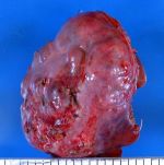
- Biopsy: Sampling cells and tissues for microscopic examination for signs
of cancer. During tumor resection surgery or as a separate test, tissue
samples are collected for examination. Partial resection of the tumor using
a large gauge needle through the abdomen is another method. Pathologists
study collected samples under a microscope and check for liver tumor cells.
Various factors influence prognosis (the chance of recovery) and treatment
options.
The following factors influence prognosis (the chance of recovery).
- Whether the complete extraction of the tumor is
possible. Type of liver tumor (the hepatoblastoma or the hepatoma)
- Initially diagnosed or relapsed case
- Extremely low levels of AFP
The following factors influence prognosis of the patients.
- The characteristics of the cancer cells (appearance at observation under
the microscope.)
- After starting chemotherapy, whether the blood levels of AFP decreases
or not.
Pediatric liver tumors may be curable when the tumor is small and complete
extraction is possible. Comparing with hepatocellular carcinoma, complete
removal of hepatoblastoma is becoming more possible recently. Even in cases
of apparently completely excisable tumors the JPLT recommends surgery in
conjunction with preoperative chemotherapy. This has helped to improve
treatment results. The Europe group (SIOPEL) also obtained good results.
The stage of the pediatric liver tumor
After diagnosis of pediatric liver tumor, more tests will be carried out
in order to clarify the spread of cancer cells in the liver and metastasis
to the other parts of the body.
The process of categorizing the extent of cancer in the liver and degree
of metastasis to other parts of the body is called staging. The stage of
the cancer is determined based on the information gathered during this
process. Identifying the stage of the cancer is important in order to make
an effective treatment plan.
The staging systems for pediatric liver tumor are as follows:
- Postoperative (after surgery) staging: stage is based on how much of the
tumor remains in the body after surgery (i.e. the classification of The
Japanese Society of Pediatric Surgeons).
- Preoperative (before surgery) staging: the liver is
divided into 4 areas (Segments 4), and stage is based on how many of these 4
segments the tumor has spread to (determined by the imaging procedures such as
MRI or CT). This staging method is called PRETEXT and JPLT now uses this
classification.
The following test methods and procedures are used in the staging process:
- CT scan: See above.
- MRI (Magnetic resonance imaging): See above.
- Ultrasound examination: See above.
- Biopsy: See above.
- Surgery: Tumors are surgically excised. Tissue
extracted during surgery is examined by pathologists.
JPLT uses the following stage classifications for preoperative pediatric
liver cancer.
PRETEXT Stage I
On Stage I, the tumor can be seen in only one section
out of 4 hepatic segments.
PRETEXT Stage II
IOn Stage II, the tumor can be seen in two adjoining segments out of 4
hepatic segments.
PRETEXT Stage III
IOn Stage II, the tumor can be seen in two adjoining segments out of 4
hepatic segments
PRETEXT Stage IV
On Stage IV, the tumor can be seen in all 4 hepatic segments.
Recurrent pediatric liver tumor
Recurrent pediatric liver tumor is cancer which has deteriorated again
(relapse) after treatment. Relapse can happen in the liver and also in
other parts of body.
Treatment Options
There are various treatments for pediatric liver tumors.
Various treatments are available for pediatric liver tumor patients. The
options are standard treatment (treatment method which is currently in
general use) and the ones currently being tested in clinical trials. Clinical
trials for treatment methods are research studies aiming at improvement
of treatment methods currently in use or information gathering for new
cancer treatments. New treatment becomes standard, only after being verified
as having excellent results by means of several clinical trials.
The incidence of childhood cancer is low, therefore, when pediatric liver
tumor has been diagnosed all patients are seriously urged to consider joining
clinical trials wherever possible. Clinical trials have been performed
all over Japan. If you wish to join this trial, please contact your nearest
JPLT participating facility. The best way to determine which treatment
is most suitable for each child is for medical professionals to work closely
together with family members.
For the treatment of pediatric liver tumor,
treatment plans need to be created for each patient by a team comprised of
several doctors who are skillful in the treatment of this rare childhood
cancer.
Treatment of this disease is led by pediatric oncologists (childhood cancer
specialists.) Pediatric oncologists may occasionally consult with pediatricians
who specialize in this specific medical field and other pediatricians who
are well versed in the treatment of pediatric liver tumor patients. Participation
of pediatric surgeons with experience in liver tumor excision is also important.
In addition to this, the following medical specialists and experts may
participate in treatment:
- Radiation oncologists
- Pediatric nurse specialists
- Rehabilitation specialists
- Psychologists
- Social workers
The following are used as a standard treatment:
Surgery
If possible, the cancer will be surgically removed.
- Partial hepatectomy: Surgery which resects the part which is invaded by
hepatic tumors. There are several kinds of excision such as wedge-shaped
excision of tissue (surgical removal of a wedge-shaped portion of tissue:
we do not recommend this method due to high possibility of relapse), hepatic
lobe, hepatic segment excision (the liver is divided into 4 segments and
right and left lobes) among others.
- Total hepatectomy and liver transplant: Treatment
which resects the whole liver and replaces it with a healthy liver offered by a
donor. A liver transplant can be performed when the tumor has not spread outside
the liver and also when you have a liver donor. While you have to wait for the
offer of a liver, the other treatments will be performed as necessary. In the
West, research into cadaveric liver transplant shows good results. However the
main method in Japan, living donor liver transplant, only has been covered by insurance
since 2010.
- Resection of metastasis: Surgery which resects metastasis outside of the
liver (in surrounding tissue, lung, brain and so forth).
In a large number of cases, preoperative
chemotherapy is used in order to shrink the tumor for easier resection and to
avoid the scatter of tumor cells at surgery. This kind of treatment is called
neoadjuvant therapy. In most of cases, postoperative chemotherapy is used to kill
any residual tumor cells, even if surgeons were able to remove all visible
tumors during surgery.
Chemotherapy
Chemotherapy is treatment using medicines to
prevent tumor growth by killing cancer cells and obstructing the cell division
of cancer cells. In case chemotherapy is administered orally (by mouth) or by
intravenous or intramuscular injection, medicine enters the bloodstream and
reaches cancer cells throughout body (systemic chemotherapy). Although, in most
cases systemic chemotherapy is used, there is a treatment called TACE which
injects locally embolic agents (localized chemotherapy) and chemotherapy agents
into an artery which flows into the liver. This hepatic artery treatment (the main
artery which provides the liver with blood containing oxygen) is one kind of
regional chemotherapy and it is used for pediatric liver tumor treatment. In
this treatment neoplastic drugs are injected into the hepatic artery through a catheter
(small tube). The medicine contains a substance which embolizes the artery and
it blocks the blood supply to the tumor. Consequently, most of the antineoplastic
agents stick around the tumor and less antineoplastic agents spread to other
parts of the body. Consequently, it also shuts out oxygen and nutrition which
promote tumor growth. Blood supply for the liver is maintained by blood flowing
through the portal vein from the stomach and the small intestine.
Treatment using multiple antineoplastic agents is
called combination chemotherapy. The type of chemotherapy implemented depends
on the type and stage of the tumor.
Radiation therapy
Radiation therapy uses radiation such as high
energy X-ray to kill cancer cells. There are two kinds of radiation treatment.
External radiation treatment irradiates cancer from outside of the body.
Internal radiation treatment places radioactive substances directly inside or
around the tumor using needles, seeds, wired and catheters. The type of
radiation therapy implemented depends on the type and stage of the tumor.
|